Anterior Exposure for Spine Surgery
The complex pathology of the spine requires a multitude of surgical solutions. The anterior approach of the spine has been around for many years but in the past was done through large incisions that oftentimes resulted in a difficult recovery and less than optimal outcomes. The progress in technology and good results with the anterior lumbar interbody fusion (ALIF) and FDA approval of the artificial disc revived the anterior lumbar spine surgery. In order for this to be successful, the surgical technique for exposure of the spine was significantly improved and nowadays, in expert hands, is done through small incisions with minimal complications.
The decision to perform an anterior spine procedure is made by the spine surgeon but these surgeries are performed by a team of an access surgeon and a spine surgeon. It is critical for the success of surgery that the spine is exposed safely and quickly to avoid injuries to the blood vessels, nerves, ureters and bowels that are in front of the spine. This is the part of the surgery done by the access surgeon.
In order to understand how this surgery is done, let’s review the anatomy of the spine.
Looking from the front, the spine appears like a column of blocks (vertebral bodies) stacked one on top of the other. Between the vertebrae there are disks and a complex system of joints which allow movement of each vertebra. The spine is covered in front by multiple structures.
All of these important structures can be grouped into 3 categories:
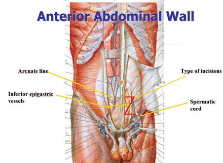
Anterior abdominal wall

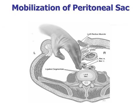
Abdominal sac (peritoneal sac) containing bowel and other organs

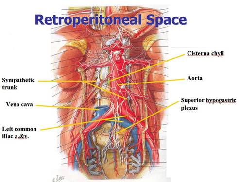
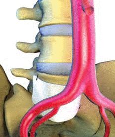
Retroperitoneal structures: the largest blood vessels of the body also called the great vessels (aorta and vena cava), blood vessels going to the legs (iliac vessels), various nerves and the tubes carrying from the kidneys to the urinary bladder (ureters)

The job of the access surgeon is to navigate between all of these structures and to expose the spine.
The surgery is done in three steps:
- Step 1: The exposure of the spine (performed by the access surgeon)
- Step 2: The spinal surgery (performed by the spine surgeon)
- Step 3: Confirmation of lack of injuries and closure of the abdomen (performed by the access surgeon)
The access surgeon begins the first step of the surgery by placing a small incision in the abdomen (for a single vertebral this is usually 2-3 inches long). The location of the incision depends on the vertebral level that needs to be exposed. This incision is longer if multiple vertebral levels need to be exposed.
The abdominal muscles are split and not cut, sparing the nerves and allowing for quicker recovery. Behind the abdominal muscles is the peritoneal sac which is moved together with the ureters, allowing entrance into the retroperitoneal space. Next the access surgeon performs the most delicate part of the procedure, wherein he moves the blood vessels and nerves away from the spine. Once this is accomplished, a retractor is placed which keeps all of these structures away from the spine while the spine surgeon performs his part of surgery.
The second part of the surgery is performed by the spine surgeon. Depending on the treatment required this could involve fusion of the spine, removal of a tumor, repair of fractures or replacing the natural disc with an artificial disc.
Once the spine surgeon finishes, the third part of the surgery is performed by the access surgeon. His objectives at this point are to insure there are no injuries and to close the abdomen. In addition, many times an adhesive barrier (Vessel Guard Patch) is placed in front of the spine to decrease the probability of scar tissue forming between the vessels and the spine. This step is done to reduce the risk of injury of the vessels in case a re-operation may be necessary.
 The procedure described above is for treatment of the lumbar spine. If, instead, surgery is to be performed on the thoracic spine, a similar access will be accomplished through the chest.
There are cases in which the spine cannot be safely exposed from the front:
- Patients with a history of prior surgeries in the retroperitoneal space
- Patients with a history of vascular surgeries involving placement of artificial vascular grafts or stents
- Patients with severely calcified vessels
- Patients with aneurysms of the aorta or iliac arteries
- Patients with morbid obesity
Possible complications after anterior exposure of the spine include:
- Infection
- Bleeding that could be very serious
- Bowel, ureter and/or bladder injury
- Spleen or kidney injury
- Retroperitoneal fluid collection
- Cord ischemia with paraplegia
- Swelling of the legs
- Blood clots in the vessels
- Warm leg
- Retrograde ejaculation and possible irreversible sterility
- Testicular pain
 Surgical Technique for sutured fixation
of Gore® Preclude® Vessel Guard Surgical Technique for sutured fixation
of Gore® Preclude® Vessel Guard
All the Patient Forms are Adobe PDF files and require an Adobe Acrobat Reader If you do not have a copy, you can obtain a free copy of the Reader from the Adobe site. Click on the Adobe logo below go download the Adobe Reader.

|